

Picture of the Month Archive
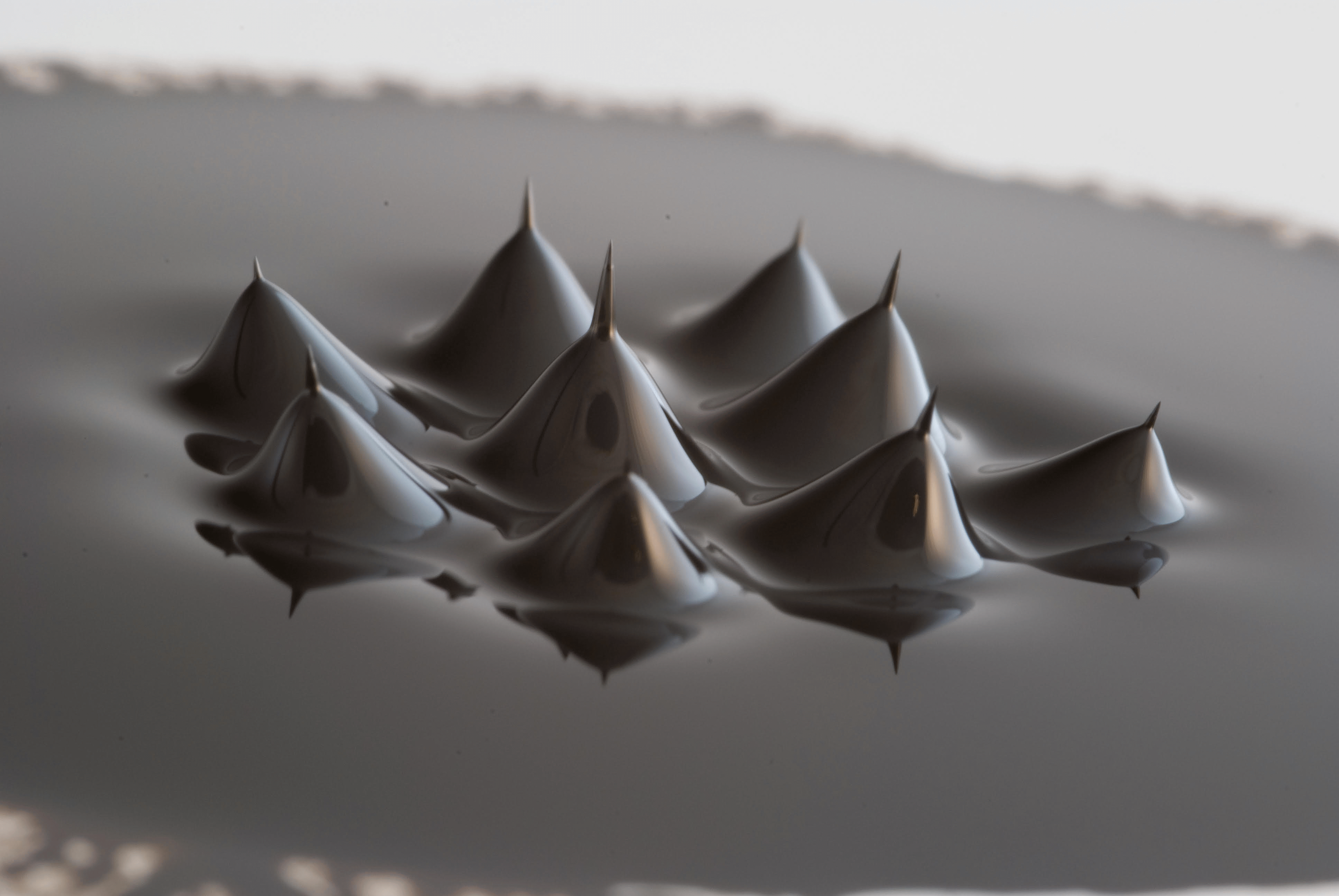
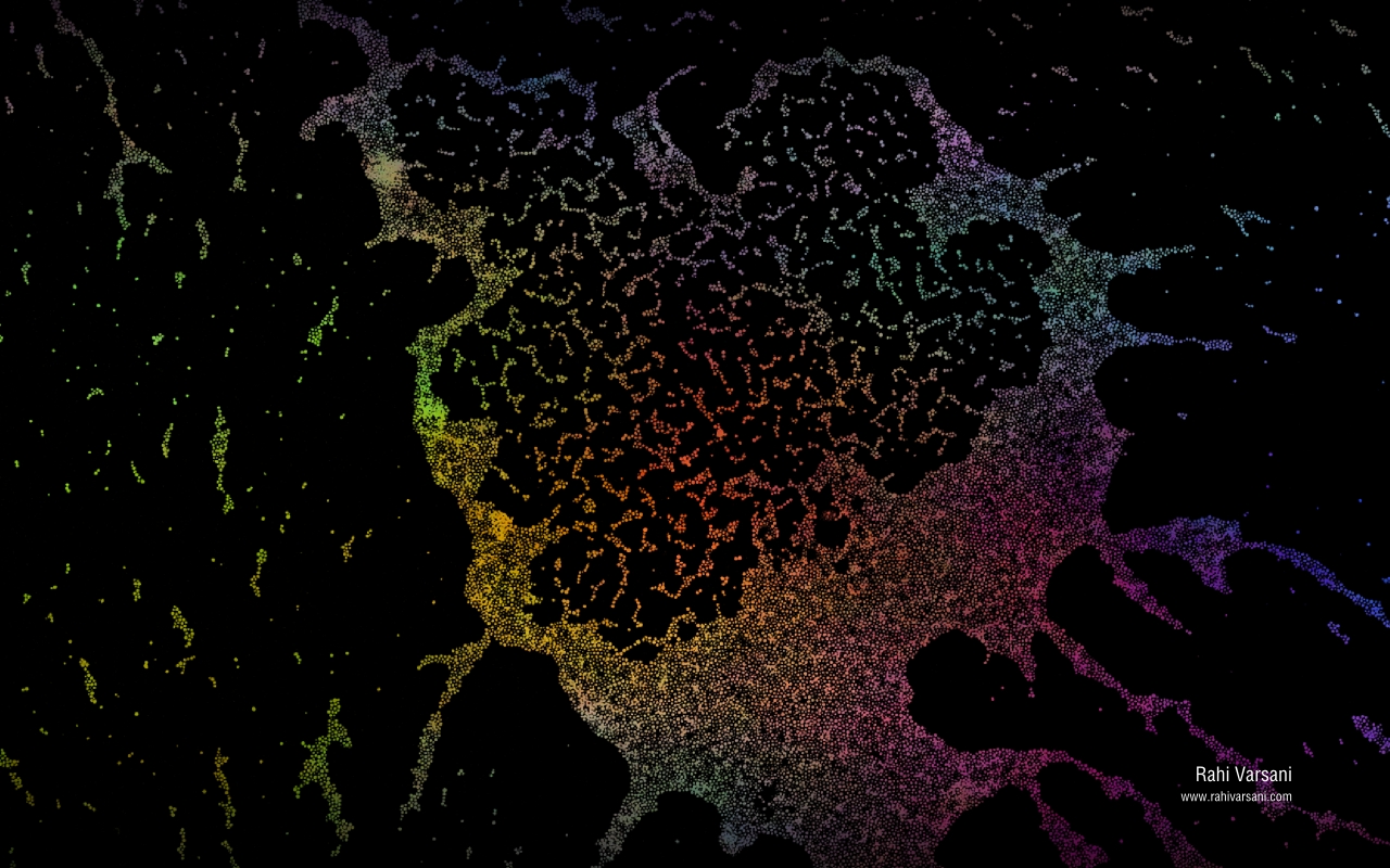
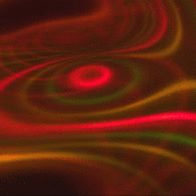
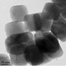
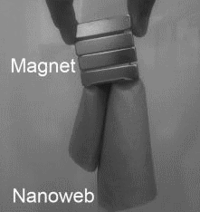
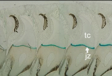
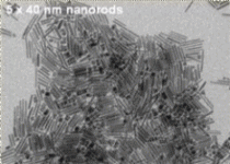
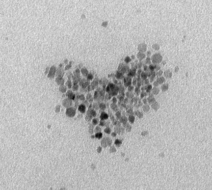
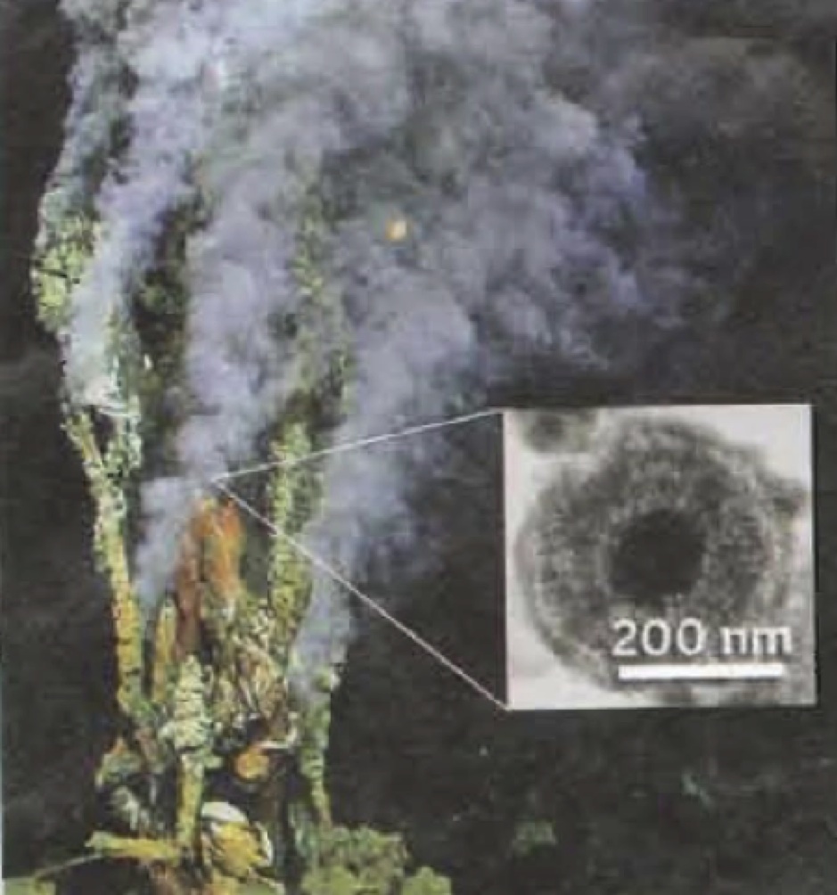
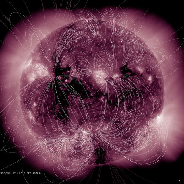
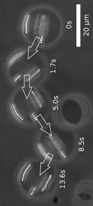
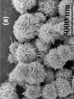
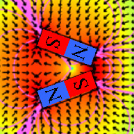
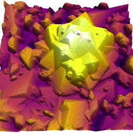
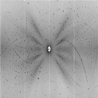
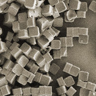
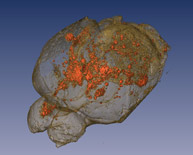
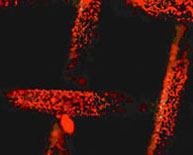
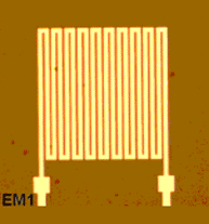
Photo of the Month
September 2017
September 2017
Beautiful ferrofluid, with a curious and striking 'peak-on-a-peak' effect. Submitted by Quentin Pankhurst.
Photo of the Month
December 2014
December 2014
Adsorbing nominal amounts of magnetite nanoparticles onto the surface of stiff, anisotropic ceramic particles like alumina enables the fast, parallel control of inorganic architectures with weak, scalable magnetic fields. This ultra-high magnetic response has been exploited to produce complex filler architectures in a new family of advanced composites. For more info, contact Randall Erb. To see the movie, click here.
Photo of the Month
January 2014
January 2014
A magnetic map of a grain of titanomagnetite from a Mount Saint Helens ash flow. Image by IRM's most recent graduate student Evgeniya Khakhalova.
Photo of the Month
November 2013
November 2013
Interesting NiCu nanowires made in the lab of S. Thongmee [Maleak N et al. (2013). J Magn Mag Mat, [http://dx.doi.org/10.1016/j.jmmm.2013.10.054].
Photo of the Month
December 2012
December 2012
EM Image of the self-assembly of magnetic iron-oxide nanoparticles. Rahi Vasani at the University of Western Australia (UWA) let a droplet with more than a trillion nanoparticles dry on a film. The high surface tension drew particles together into this beautiful picture which makes a cool computer background!
Photo of the Month
November 2012
November 2012
What you see here is art made with a ferrofluid lens. If you want one, check our Michael Snyder's website at www.revolution-labs.com.
Photo of the Month
October 2012
October 2012
Microfluidic train of magnetic droplets (33 nL) in a thermally insulating oil to confine the heat produced by RF hyperthermia. D. Habault et al. to appear in IEEE Trans Mag
http://arxiv.org/abs/1209.5249
http://arxiv.org/abs/1209.5249
Photo of the Month
September 2012
September 2012
Nice TEMs of carbon encapsulated iron oxide/iron carbide nanocomposites for hyperthermia. Sharma M, Mantri S, Bahadur D (2012). JMMM 324, 3975-3980.
Photo of the Month
June 2012
June 2012
Magnets usually attract magnetic materials, however, two or more magnets can be arranged to generate a push force as can be seen in the video where a steel ball bearing is levitated against gravity. This movie is from Azeem Sarwar, and Ben Shapiro at the University of Maryland College Park, USA (2012).
Photo of the Month
May 2012
May 2012
Nanoparticles are embedded into electrospun nanofibres and can then be used to make magnetic clothes! Sung, Ahn, Kang, JMMM 324, 916 (2012).
Photo of the Month
April 2012
April 2012
Have you heard of chitons, these marine molluscs with a shell? Interestingly, they grow iron teeth, so can be detected magnetically. The biomineralization of chitons was researched by Jeremy Shaw, Martin Saunders et al. at the University of Western Australia in Perth.
Photo of the Month
March 2012
March 2012
Variant shape growth of nanoparticles of metallic Fe-Pt, Fe-Pd and Fe-Pt-Pd alloys, Nguyen T. K. Thanh et al.
Photo of the Month
February 2012
February 2012
Happy Valentine's Day from the magnetite world. Submitted by Lucía Gutiérrez, UWA, Perth, Australia and ICMM, Madrid, Spain.
Photo of the Month
September 2011
September 2011
Deep-Sea Vents Dispense Nutritious Pyrite Nanoparticles! Click HERE for the full story
Photo of the Month
March 2011
March 2011
Magnetic fields around the sun.
Photo of the Month
November 2010
November 2010
Well coated superparamagnetic nanoparticles make beautiful ferrofluids (right), while less stable ones (left) agglomerate in high salt concentrations (e.g., blood!) under the influence of an applied magnetic field. This movie is from Prof Etelka Tombacz at the University of Szeged in Hungary (2010).
Photo of the Month
October 2010
October 2010
Separation and subsequent culturing of MCF-7 breast cancer cells on self-assembled protein-coated magnetic beads in a microfluidic chip. By Sivagnanam and Gijs et al.
Photo of the Month
September 2010
September 2010
Micrograph of a vesicle which includes about 20 superparamagnetic
beads being chained up. Such particles might be useful for micromixing. By Franke et al 2009.
beads being chained up. Such particles might be useful for micromixing. By Franke et al 2009.
Photo of the Month
August 2010
August 2010
SEM (a,b,c) and TEM micrographs (d,e) of superparamagnetic nanostructures of %u03B1-Fe2O3 taken by Cao et al 2009..
Photo of the Month
July 2010
July 2010
| With a smart arrangement of more than one magnet, microparticles can be pushed (!) away, as shown here by Benjamin Shapiro, Ken Dormer, Roland Probst and Isaac Rutel. Click for the full detailed image. |
Photo of the Month
June 2010
June 2010
Interesting scan of a magnetite crystal.
Photo of the Month
May 2010
May 2010
Levitation of a chaperoned droplet with a magnet. The adhesive and magnetic forces in the porous Si chaperones are sufficient to allow pick up and placement of a 2%u20134 mm diameter aqueous droplet. Dorvee J, Sailor M, Miskelly G (2008). Dalton Trans 6, 721-730.
Photo of the Month
April 2010
April 2010
Neutron lauegram of the first chemically and magnetically chiral molecular magnet (Clara Gonzalez and Fernando Palacio).
Photo of the Month
March 2010
March 2010
Transportation of magnetic particles in a staircase pattern of magnetic cylinders (2x6x0.1 µm) in an applied rotating magnetic field. Movie by Klas Gunnarsson et al., Adv Mater 2005, 17, 1730-1734
Photo of the Month
February 2010
February 2010
Magnetic 2 µm polymer cubes were made using the PRINT technology (particle replication in non-wetting templates) by K.P. Herlihy and J.M. DeSimone, Proc. SPIE 6517, 651737 (2007).
Photo of the Month
January 2010
January 2010
Many magnetic iron oxides have distinct coloures, as shown in these colour tables from Cornell & Schwertmann 2003.
Photo of the Month
December 2009
December 2009
3D MRI reconstruction of mouse brain following injection of magnetically labeled neural stem cells shows widespread dissemination of cells throughout the brain (courtesy of Piotr Walczak and Jeff Bulte).
Photo of the Month
June 2009
June 2009
Magnetic separation on a chip is nicely shown by this movie that you see after clicking on the chip! High field gradients along the magnetizable strips efficiently separate the particles. Courtesy of Sang-Hyun Oh, University of Minnesota.
Photo of the Month
December 2008
December 2008
Incorporating magnetic nanoparticles into a spider silk "beam" led to a magnetically responsive structure (magnet held above the silk) (Jiamei Bai et al, University of British Columbia, Vancouver, Canada).
Photo of the Month
December 2007
December 2007
The magnetic stent, presented at our conference in Lyon, demonstrates that magnetic microspheres carrying therapeutic substances can be delivered to magnetized cardiovascular implants by simple intra-arterial injection (Benjamin Yellen et al 2004).
Photo of the Month
December 2006
December 2006
These pictures are on our 2006 conference poster. They show magnetic coils on a silicon chip and were developed by Dr. Qasem Ramadan, Nanyang University, Singapore.