

The 13th Ferrofluid Workshop is Coming Up !
July 23, 2013
 This year's and now already 13th Ferrofluid Workshop in Benediktbeuern, Germany will take place from September 25-27, 2013.
Check out the details here:
http://www.mfd.mw.tu-dresden.de/ffworkshop/
Tannic Acid and Fe(III) Form Special Coating on Almost Any Substrate
July 19, 2013
 A team led by Frank Caruso at the University of Melbourne has developed a simple one-pot recipe for a coating made of only Fe(III) and tannic acid, a polyphenol found in wood that is perhaps best known for improving the flavor of wood-casket-aged red wine. The coating is unusually versatile. It can cover all manner of nano- and microscopic objects, including
gold nanoparticles, calcium carbonate and silicon dioxide particles, and bacteria, regardless of whether the object to be coated is positively charged, negatively charged, or neutral ( Science 2013, DOI:10.1126/ science.1237265). The discovery is patentpending.
Since both Fe(III) and tannic acid are generally regarded as safe by regulators and are already used in food and biomedical applications, the coating could find uses right away in areas as diverse as drug delivery and corrosion protection, comments Christopher W.
Bielawski , a chemist at the University of Texas, Austin.
“This is going to make a big impact mainly because it is so simple,” he adds. “Many will wish that they had thought of it first.”
“The coating’s pH sensitivity is the really exciting aspect,” comments Phillip B. Messersmith , a biomedical engineer at Northwestern University. The coating forms above pH 6, but in more acidic environments it falls apart, revealing or releasing the contents of objects within it. One could envision using the coating as a way to deliver a drug to a cell’s acidic lysosome or endosome and then having the contents released in the organelle’s low-pH environment, he says. Researchers use “tannic acid” to describe a family of molecules that contain a central glucose with one to five polygalloyl groups of varying lengths that emanate from the sugar base. Every Fe(III) atom can coordinate three pairs of hydroxyl groups found on tannic acid and thus can complex up to three different tannic acid molecules. Meanwhile, the abundance of hydroxyl groups in tannic acid means that each tannic acid molecule can complex up to a dozen or so iron atoms. The result is a cross-linked coating that is about 10 nm thick, Caruso says.
The technique seems like it could be easily expanded, Bielawski says. “There are a lot of other polyphenols out there besides tannic acid, so there’s considerable potential for changing the surface chemistries of the coating and creating new applications.”
Improve Energy Conversion with the Help of Magnetic Nanoparticles
July 15, 2013
 AN OBSTACLE to developing low-cost energy conversion devices is understanding why small differences in materials can strongly affect performance. Figuring out the relationship between the structure and function of an electrode material, for example, is especially challenging for nanoparticle aggregates.
Researchers have now developed an analytical procedure to scrutinize material structure and function with nanometer resolution across micrometer distances in such aggregates. The technique enables them to design a better electrode for solar energy conversion. The group used transmission electron microscopy (TEM) to image the spatial distribution and orientation of nanocrystals within aggregates.
Then, by using a conducting atomic force microscopy method, they correlated the TEM information with the pathways that electrons follow as they move through the material. They used the combo method to probe electron transport in various nanoparticle-based iron oxide electrodes. The electron-transport proficiency of this cheap material makes it useful for generating hydrogen via light-driven water splitting. Although the electrodes they studied were similar, some worked well, while others did not.
The key finding of the team, which includes Scott C. Warren of the University of North Carolina, Chapel Hill, is that the relative orientation of adjacent nanoparticles greatly affects charge transport. A small orientation mismatch is alright, but a large mismatch results in electrical barriers that block current flow between adjacent grains. The upshot is that by identifying “winning” crystal orientations and tailoring the preparation method to favor them throughout the electrode, the group made a device that achieves a record-setting photocurrent for this class of materials ( Nat. Mater. 2013, DOI: 10.1038/nmat3684).
“These results represent an important step forward in developing nanostructured materials for next-generation energy conversion devices,” says solar-fuel specialist Roel van de Krol of Technical University of Berlin. Boston College chemist Dunwei Wang adds that the study helps explain why some nanostructures function better than others. “This work will be of value to photoelectrochemistry studies in general,” he adds. — MITCH JACOBY, C&EN
Conversion of Carbon Dioxide to Chemically Useful Products with the Help of Magnetic Nanoparticles
July 01, 2013
.gif) CO2 gets a lot of attention - mostly negative - as a greenhouse gas. But if CO2 could be converted in a cost-effective manner to valuable products, the ubiquitous small molecule so often reviled for its role in climate change might start to lose its bad rap.
CO2 gets a lot of attention - mostly negative - as a greenhouse gas. But if CO2 could be converted in a cost-effective manner to valuable products, the ubiquitous small molecule so often reviled for its role in climate change might start to lose its bad rap.
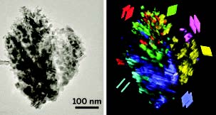
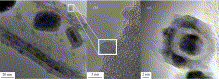
Working to make this happen is Siglinda Perathoner at the University of Messina in Italy. As a catalysis specialist, she and her co-workers have developed methods for preparing catalytic electrodes that consist of iron nanoparticles supported on carbon nanotubes (Fe/CNTs). The picture above shows the overall view, while the picture below zooms in (with HRTEM) onto the magnetic nanoparticles both inside and on the outside of the CNTs. In contrast to the liquid-phase reactions occurring in the electrocatalytic cells used by some researchers, their system converts CO2 in the gas phase. In this way, she sidesteps the need to separate products from a liquid mixture and avoids potential limitations in the amounts of the starting material that can be dissolved in aqueous solution.
 Peratoner acknowledges that nanotubes decorated with Pt nanoparticles are more stable and in some cases more active catalysts than Fe/CNTs. Yet Fe/CNTs, especially ones doped with nitrogen, are highly active—for example, in producing isopropyl alcohol—and are especially attractive because of their low cost. For more information, check out her recent paper here.
Magnetic Nano-Stirbars
June 29, 2013
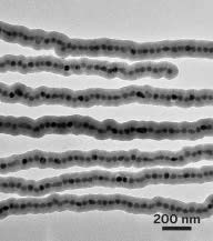 Researchers who want to combine tiny volumes of liquid have few simple options when it comes to mixing for lab-on-a-chip applications or microliter bioassays. Passive diffusion is slow, and violent stirring can break droplets apart instead of mixing them together.Thanks to a team in Singapore, researchers can now reach for the world’s smallest magnetic stir bars. At just 17 μm long and 75 nm to 1.4 μm thick, these super small stir bars can mix as little as 4 pL of liquid (Angew. Chem. Int. Ed. 2013, DOI:10.1002/anie.201303249). A team at Nanyang Technological University led by Hongyu Chen created the stir bars from oleic acid-stabilized Fe3O4 nanoparticles that are 40 nm in diameter. They modified the nanoparticles with citric acid to render them water-soluble and dissolved them.
The team then used a magnet to align the nanoparticles and gave them a silica coating to ensure they stayed straight and rigid. They centrifuged the mixture to purify the stir bars, which they dispersed in solutions of various concentrations for stirring. While stirring, the nanosized stir bars remain suspended in solution indefinitely. And removing them is as simple as placing a stationary magnet beneath the droplet and letting the stir bars settle out of solution over the course of about five minutes.
Preventing Food-Borne Illness
June 29, 2013As recently highlighted at the ACS (American Chemical Society) meeting, nanomaterial and especially magnetic-particle-based sensors might one day help to prevent food-borne illness by quickly detecting bacteria such as Listeria on food and in the water used to wash food.
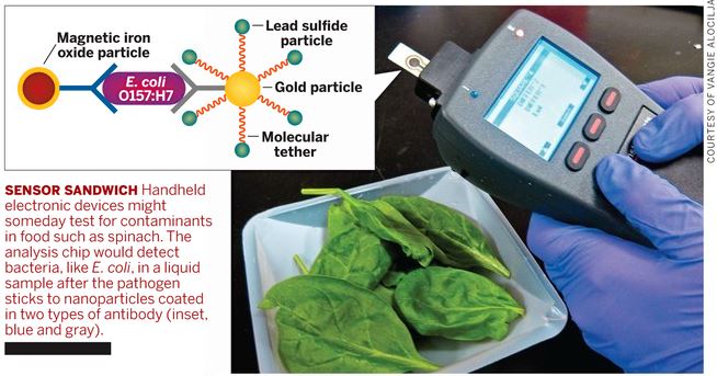
For more information, check out the entire article here.
MagForce AG Receives Approval to Start the Post-Marketing Study in Glioblastoma with NanoTherm® Therapy
June 26, 2013
.jpg) MagForce AG (Frankfurt, XETRA: MF6), a leading medical technology company in the field of nanomedicine in oncology, announced today that it has received approval from the German Federal Institute for Drugs and Medical Devices (BfArM) to start the post-marketing clinical study in recurrent glioblastoma. The approval of the ethics committee was previously granted end of December 2012 and the Company will now proceed to install the required NanoActivators™ in the treatment centers and preparing for the clinical study initiation.
MagForce AG (Frankfurt, XETRA: MF6), a leading medical technology company in the field of nanomedicine in oncology, announced today that it has received approval from the German Federal Institute for Drugs and Medical Devices (BfArM) to start the post-marketing clinical study in recurrent glioblastoma. The approval of the ethics committee was previously granted end of December 2012 and the Company will now proceed to install the required NanoActivators™ in the treatment centers and preparing for the clinical study initiation.
The trial is an open-label, randomized, controlled study to determine the efficacy and safety of NanoTherm® monotherapy alone and in combination with radiotherapy versus radiotherapy alone in up to 280 glioblastoma patients. It will be conducted in about 15 centers in Germany and will be initially started in five leading centers, including the University Hospitals Berlin, Duesseldorf, Giessen, Cologne and Muenster. Together with a steering committee of leading experts in neurooncology, MagForce has designed a study protocol to support and supplement the results of a previous study, which led to the approval of the NanoTherm® Therapy and its medical devices in brain cancer. In addition, the application procedure of the NanoTherm® particles into the tumor should be optimized. This study is also designed to comply with the current guidelines for the development of medicinal products.
"We are excited about the BfArM approval that now triggers the start of our post-marketing clinical study, which we have carefully prepared over the last few months in collaboration with leading neurosurgeons, neurooncologists, and radiotherapists. With this trial we intend to provide validation of the available results with our NanoTherm® Therapy involving leading medical experts in the field of neurooncology, the key opinion leaders in the application of this technology", Prof Dr Hoda Tawfik, COO and co- CEO of MagForce, commented.
For more information, check out MagForce's website at
http://www.magforce.de/en/studien/uebersicht.html
Iron and Magnetism in Schistosome Eggshells
May 28, 2013
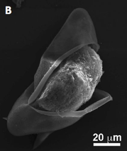 Quite interesting research from Tim St. Pierre's lab in Perth, Western Australia was recently published in PLOS Neglected Tropical Diseases. Approximately 200 million people in the world are infected with schistosomes. Diagnosis of schistosomiasis is often difficult and improved diagnostic methods are urgently needed to evaluate the success of elimination programs. Recently, a magnetic fractionation method for isolation of parasite eggs from feces was described, which uses magnetic microspheres to form parasite egg – magnetic microsphere conjugates. In an elegant investigation, Stephan Karl and Lucia Gutierrez followed up on the unexplained mechanism of formation of the conjugates.
The researchers determined the magnetic properties of the eggs, studied the motion of eggs and egg-microsphere conjugates in magnetic fields and determined species specific affinity of parasite eggs to magnetic microspheres. Iron is predominantly localized in pores in the eggshell. Beautiful electron microscopy pictures and investigations suggest that the interaction of magnetic microspheres and parasite eggs is unlikely to be magnetic in origin. Instead, the filamentous surface of the eggshells may be important in facilitating the binding. Modification of microsphere surface properties may therefore be a way to optimize magnetic fractionation of parasite eggs.
For more information, check out our Archives.
September 2017
Search this site with the power of